flow cytometry mds
Submit 1 - 3 mL if bone marrow in a special bone marrow collection tube available by calling the NorDx Flow Cytometry Lab 207-396-7912. Run sticky samples at high flow rates with a system that is less sensitive to clogging.

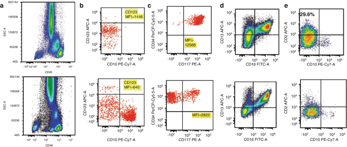
Multi Parameter Flow Cytometric Analysis Showing Cytoplasmic Download Scientific Diagram
Flow cytometric FCM approaches have been described.

. Analysis by flow cytometry FC of bone marrow cells has been introduced as an important co-criterion in the diagnosis of MDS. MDS is often referred to as a bone marrow failure disordercommonly found in the aging population. Ad Confirm verify and optimize your gated cell populations with real-time image tracking.
We sought to refine the FCM approach by using peripheral blood PB to create a clinically useful tool for the diagnosis of MDS. Contains Lysing Solution and Fixation Permeabilization Wash Buffers For Flow Cytometry. Flow cytometry FCM analysis can be an important tool and provide diagnostic clarity in cases where patients with cytopenia are suspected of having myelodysplastic syndrome MDS according to a.
Ad Buy Intracellular Flow Cytometry Reagents Conjugated Monoclonal Antibodies. Protocol for a Diagnostic Accuracy Study MPO-MDS-Valid The safety and scientific validity of this study is the responsibility of the study sponsor and investigators. These include abnormalities in the quantity and phenotype of blasts the.
However the value of bone marrow immunophenotyping in MDS remains unclear due to the variability in detected abnormalities. Multiparameter flow cytometry MFC is upcoming in MDS diagnostic work up comparability and investigator experiences are critical. In suspected MDS patients multi-parameter flow cytometry can aid in establishing diagnosis risk stratification and choice of therapy.
Guidelines recommend flow cytometric analysis as part of the diagnostic work-up of cytopenic cases suspected for myelodysplastic syndromes MDS Porwit et al 2014. Recently a flow cytometric score FCM-score was published capable of discriminating low-grade MDS from non-clonal cytopenias Della Porta et al 2012. Van de Loosdrecht et al 2022Many reports on flow cytometry FCM in MDS focus on examples of aberrant immunophenotypic features without.
Myeloid nuclear differentiation antigen MNDA in myelomonocytic cells might be expressed more weakly in patients with MDS. Flow cytometric FCM approaches have been described. Additionally we propose an integrated diagnostic algorithm for suspected MDS.
Flow cytometry assessment of MDS Abnormal flow cytometry patterns predict 4MDS with good sensitivity and specificity WHO 2016 and ELN guidelines do not permit a diagnosis of MDS solely based on flow cytometry 10 onsidered supportive of. Porwit et al 2022. Jaffe MD in Hematopathology 2017 Flow Cytometry Abnormalities.
Mayo Approach The results of our studies are interpreted as normal atypical or aberrant. This review addresses the developments and future directions of multi-parameter flow cytometry scores in MDS. Ad Buy Intracellular Flow Cytometry Reagents Conjugated Monoclonal Antibodies.
Forward promptly at ambient temperature only. Our sensitivity and specificity are approximately 70 and 95 respectively. As research has shown that flow cytometry is capable of diagnosing reactive and clonal proliferations of bone marrow hematopoietic cells the method has been explored as a potential diagnostic tool.
Myelodysplastic syndromes MDS are classified by the WHO as myeloid neoplasms and are characterized by cytopenia and dysplasia in one or more myeloid cell lines. We sought to refine the FCM approach by using peripheral blood PB to create a clinically useful tool for the diagnosis of MDS. They can be some of the most difficult diagnoses to make in both developed and developing countries.
The analysis of MNDA may thus improve diagnostic capabilities of MFC in MDS. Valent Orazi et al 2017. The amount of tissue needed is dependent on the cellularity of the specimen.
Myelodysplastic syndromes MDS are a group of hematological disorders presenting with one or more clonal cytopenias significant dyspoiesis and increased blasts in some cases. Generally a 2-10 mm section of fresh tissue is adequate. However the value of bone marrow immunophenotyping in MDS remains unclear due to the variability in detected abnormalities.
Recently a flow cytometric score FCM-score was published capable of discriminating low-grade MDS from non-clonal cytopenias Della Porta et al 2012. Myelodysplastic syndromes MDS are classified by the WHO as myeloid neoplasms and are characterized by cytopenia and dysplasia in one or more myeloid cell lines. Contains Lysing Solution and Fixation Permeabilization Wash Buffers For Flow Cytometry.
Myelodysplastic syndromes in a multicenter approach. Flow Cytometric Analysis of Peripheral Blood Neutrophil Myeloperoxidase Expression for Ruling Out Myelodysplastic Syndromes. MDS hematopoietic cells exhibit recurring quantitative and qualitative abnormalities in antigen expression and maturation patterns that can be interrogated by multiparameter flow cytometry immunophenotyping.
Over the past few years significant progress has been made in the FCM field concerning technical issues including software and hardware and pre-analytical procedures. Flow cytometric analysis of human bone Working Party on Flow Cytometry in MDS. 2 FC can identify specific aberrations on both immature and maturing.
Flow Cytometry In MDS. Implementation of flow cytometry in the diagnostic work-up of Hematol Oncol 2008. Flow cytometry FCM aids the diagnosis and prognostic stratification of patients with suspected or confirmed myelodysplastic syndrome MDS.
We decided to err on the side of caution and sacrifice some sensitivity in order to make sure that we dont overcall aberrant findings. Ad Confirm verify and optimize your gated cell populations with real-time image tracking. Myelodysplastic Syndromes MDS are a group of diverse bone marrow disorders in which the bone marrow does not produce enough healthy blood cells.
Run sticky samples at high flow rates with a system that is less sensitive to clogging. Report from the Dutch 55 Loken MR Shah VO Dattilio KL Civin CI.

A Series Of Case Studies Illustrating The Role Of Flow Cytometry In The Diagnostic Work Up Of Myelodysplastic Syndromes Westers Cytometry Part B Clinical Cytometry Wiley Online Library

Comparison Of Flow Cytometry With Other Modalities In The Diagnosis Of Myelodysplastic Syndrome Pembroke International Journal Of Laboratory Hematology Wiley Online Library

The Flow Cytometry Myeloid Progenitor Count A Reproducible Parameter For Diagnosis And Prognosis Of Myelodysplastic Syndromes Johansson Cytometry Part B Clinical Cytometry Wiley Online Library

Morphological Flow Cytometry And Cytogenetic Diagnosis Of Mds Springerlink

Gating Strategy For The Determination Of The Different Cell Populations Download Scientific Diagram

Five Color Flow Cytometric Analysis Of The Progression From Blasts To Download Scientific Diagram

The Flow Cytometry Myeloid Progenitor Count A Reproducible Parameter For Diagnosis And Prognosis Of Myelodysplastic Syndromes Johansson Cytometry Part B Clinical Cytometry Wiley Online Library

Five Color Flow Cytometric Analysis Of The Progression From Blasts To Download Scientific Diagram

Flow Cytometry Immunophenotyping In Integrated Diagnostics Of Patients With Newly Diagnosed Cytopenia One Tube 10 Color 14 Antibody Screening Panel And 3 Tube Extensive Panel For Detection Of Mds Related Features Porwit 2015

Examples Of Flow Cytometry Plots Illustrating Some Aberrant Phenotypes Download Scientific Diagram

Multiparametric Flow Cytometry Analysis Of Representative Control Bone Download Scientific Diagram

Flow Cytometry Immunophenotyping In Integrated Diagnostics Of Patients With Newly Diagnosed Cytopenia One Tube 10 Color 14 Antibody Screening Panel And 3 Tube Extensive Panel For Detection Of Mds Related Features Porwit 2015

Optimized Gating Strategy And Supporting Flow Cytometry Data For The Determination Of The Ki 67 Proliferation Index In The Diagnosis Of Myelodysplastic Syndrome Data In Brief

Dysplasia Evaluation In Myeloid Progenitor Cells Leukocytes Were Download Scientific Diagram

Eight Color Flow Cytometric Analysis Of Monocytic Antigenic Expression Download Scientific Diagram

0 Response to "flow cytometry mds"
Post a Comment